Implants · DENTALVIEW GUIDE
CBCT capacity versus your own examination
Registered CBCT count is a capacity signal, not proof that imaging is needed or interpreted well for you.
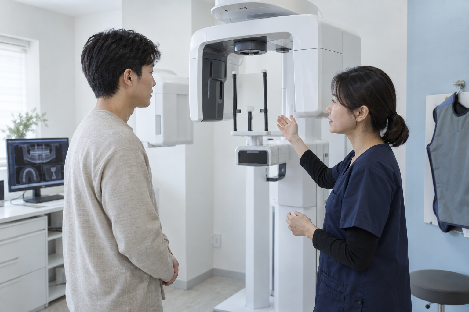
Registered CBCT count is a capacity signal, not proof that imaging is needed or interpreted well for you.
HIRA's cone-beam CT count is administrative data. Ownership does not establish your need, image adequacy or interpretation process.
For implant planning, define the question each image answers and how its findings affect position, added procedures and alternatives.
1. Separate registered count from your examination
A registered count is only a clue to equipment holdings; it does not show operation, protocol or reading quality.
Before booking, confirm availability only; assess clinical need after examination. More units do not imply better examination or outcomes.
- Before booking, confirm availability only; assess clinical need after examination.
- More units do not imply better examination or outcomes.
Official sources for this sectionHIRA medical institution detail service / KDCA National Health Information Portal: Dental implant
2. Define the question before scanning
Questions about bone dimensions, nerves, sinuses or lesions guide the field and resolution.
Ask the indication and which treatment decision the result could change. 'CT because it is an implant' is not a complete individual rationale.
- Ask the indication and which treatment decision the result could change.
- 'CT because it is an implant' is not a complete individual rationale.
Official sources for this sectionHIRA medical institution detail service / KDCA National Health Information Portal: Dental implant
3. Check field of view and prior images
Wider or repeated imaging is not automatically useful. Review the date, field and quality of existing studies.
Separate what prior images answer from why a new scan is needed. The balance of exposure and quality depends on purpose and patient factors.
- Separate what prior images answer from why a new scan is needed.
- The balance of exposure and quality depends on purpose and patient factors.
Official sources for this sectionHIRA medical institution detail service / KDCA National Health Information Portal: Dental implant
4. Ask who interprets and documents it
Imaging does not end with acquisition. Ask who reviews the target and other relevant findings and documents them.
Ask the clinician to point out key findings and record a summary. Patients should not diagnose normal or abnormal from the screen alone.
- Ask the clinician to point out key findings and record a summary.
- Patients should not diagnose normal or abnormal from the screen alone.
Official sources for this sectionHIRA medical institution detail service / KDCA National Health Information Portal: Dental implant
5. Trace findings into the treatment plan
The scan becomes useful when linked to position, angulation, dimensions, grafting and alternatives.
Write one line each for finding, plan change and remaining uncertainty. Detailed imaging does not guarantee healing or treatment outcomes.
- Write one line each for finding, plan change and remaining uncertainty.
- Detailed imaging does not guarantee healing or treatment outcomes.
Official sources for this sectionHIRA medical institution detail service / KDCA National Health Information Portal: Dental implant
A same-condition consultation table
Record the evidence, effect on your plan and unresolved condition—not only yes or no.
| Criterion | Before the visit | At consultation | Interpretation limit |
|---|---|---|---|
| Separate registered count from your examination | Before booking, confirm availability only; assess clinical need after examination. | How does the public count differ from the need for my scan? | More units do not imply better examination or outcomes. |
| Define the question before scanning | Ask the indication and which treatment decision the result could change. | What question will this scan answer, and what plan could change? | 'CT because it is an implant' is not a complete individual rationale. |
| Check field of view and prior images | Separate what prior images answer from why a new scan is needed. | What is missing from prior images, and what field is needed? | The balance of exposure and quality depends on purpose and patient factors. |
| Ask who interprets and documents it | Ask the clinician to point out key findings and record a summary. | Who interprets the scan, and where are key findings recorded? | Patients should not diagnose normal or abnormal from the screen alone. |
| Trace findings into the treatment plan | Write one line each for finding, plan change and remaining uncertainty. | How exactly did this finding change position, added care or alternatives? | Detailed imaging does not guarantee healing or treatment outcomes. |
| Cost by stage | Separate tests, placement, prosthesis and care | What triggers extra cost? | A headline price may not cover the full course |
| Clinician and follow-up | Record names and contact route | Who handles changes or urgent issues? | Institution size does not prove continuity |
What to verify
Ten questions to ask verbatim
- How does the public count differ from the need for my scan?
- What question will this scan answer, and what plan could change?
- What is missing from prior images, and what field is needed?
- Who interprets the scan, and where are key findings recorded?
- How exactly did this finding change position, added care or alternatives?
- What is confirmed now, and what waits for further tests?
- What realistic alternatives include tooth preservation, a bridge or a denture?
- Can I receive stage costs and added-cost triggers in writing?
- Who actually handles each stage, and how are handoffs managed?
- How do I distinguish expected recovery from reasons to call promptly?
FAQ
FAQ
What matters most when checking separate registered count from your examination?
A registered count is only a clue to equipment holdings; it does not show operation, protocol or reading quality. Before booking, confirm availability only; assess clinical need after examination. More units do not imply better examination or outcomes. At consultation, ask “How does the public count differ from the need for my scan?” and record both the supporting finding and anything still unresolved.
What matters most when checking define the question before scanning?
Questions about bone dimensions, nerves, sinuses or lesions guide the field and resolution. Ask the indication and which treatment decision the result could change. 'CT because it is an implant' is not a complete individual rationale. At consultation, ask “What question will this scan answer, and what plan could change?” and record both the supporting finding and anything still unresolved.
What matters most when checking check field of view and prior images?
Wider or repeated imaging is not automatically useful. Review the date, field and quality of existing studies. Separate what prior images answer from why a new scan is needed. The balance of exposure and quality depends on purpose and patient factors. At consultation, ask “What is missing from prior images, and what field is needed?” and record both the supporting finding and anything still unresolved.
What matters most when checking ask who interprets and documents it?
Imaging does not end with acquisition. Ask who reviews the target and other relevant findings and documents them. Ask the clinician to point out key findings and record a summary. Patients should not diagnose normal or abnormal from the screen alone. At consultation, ask “Who interprets the scan, and where are key findings recorded?” and record both the supporting finding and anything still unresolved.
What matters most when checking trace findings into the treatment plan?
The scan becomes useful when linked to position, angulation, dimensions, grafting and alternatives. Write one line each for finding, plan change and remaining uncertainty. Detailed imaging does not guarantee healing or treatment outcomes. At consultation, ask “How exactly did this finding change position, added care or alternatives?” and record both the supporting finding and anything still unresolved.
Can online information settle this decision?
HIRA's cone-beam CT count is administrative data. Ownership does not establish your need, image adequacy or interpretation process. Online information is a starting point for questions, not a substitute for examination and the treating clinician's explanation. Use these items as a checklist and mark missing information as unverified, not absent.
If two clinics disagree, what should I align first?
For implant planning, define the question each image answers and how its findings affect position, added procedures and alternatives. First confirm that both clinics used the same records, goals and cost scope, then document why their answers differ and which further tests each needs. Align the actual clinician and follow-up scope as well as price and time.
When should I pause and seek more information?
Questions about bone dimensions, nerves, sinuses or lesions guide the field and resolution. Pause when the supporting findings, actual clinician, total scope or problem-response plan remains unclear. Ask for the missing record before committing; only an examining clinician can judge urgency. Record when the review will be completed and the next action so necessary care is not lost during the pause.